

3. CELL CULTURES/GENETICS
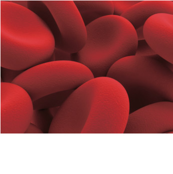
SYSTEM ABO-Rh DETERMINATION
In this experiment, students will learn the techniques used to determine the blood type and Rh.
The test procedure used in this kit is the same as used with real blood, with the only difference that this kit contains synthetic blood and synthetic antiserum eliminating any risk associated with exposure to real blood or blood products.
Each individual student has to indicate the blood group and Rh from 4 samples problems.
> Kit includes:
50 slides, 150 sticks, 12 synthetic antiserum samples, and 12 micropipettes (1.2 ml).
> You need:
All you need are included on the kit.
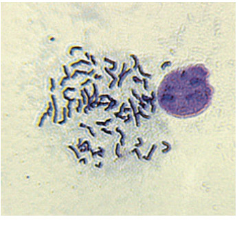
CHROMOSOME SPREAD (PRE-FIXED SLIDES)
During mitosis, each of our chromosomes is duplicated. The chromosomes are then separated during mitosis, moving to opposite ends of the cell before cell division. In this experiment, cells have been arrested during metaphase and fixed to slides, allowing students to stain and observe the condensed chromosomes.
Students will develop an understanding of karyotyping and the association of chromosomal abnormalities with diseases.
> Kit includes:
Pre-fixed chromosome spread slides, giemsa stain, mounting media, cover slips, transfer pipettes immersion troughs.
> You need:
A microscope with 400x magnification.
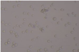
BIOLOGY AND CULTURE OF EUKARYOTIC CELLS
Cell Culture is a vital technology used in life science research and in biotechnology laboratories. The study of basic cell biology, diseases and cancer, the development and testing of new therapeutics, and the production of new drugs relies on using the techniques introduced in this experiment. Students will learn how to grow eukaryotic cells in culture, basic cell staining and how to count cells. The techniques used in these experiments will provide the student with a skill set desired in both academic research and industry.
To ensure the success of the practice IT IS NECESSARY to coordinate the day of delivery of the kit with the day of the start of the practice. To schedule the delivery date of the kit, contact the person in charge of the cell culture laboratory at the email virgili@danagen.es
> Kit includes:
Growth media, flasks, Giemsa stain, Trypan blue dye, sterile T25 flasks, sterile culture dishes, sterile large pipettes, small pipettes, cell counting chambers, Sf9 insect cells.
> You need:
Microscope and inverted microscope, Spray bottle with 70% ethanol or methanol, pipette controller.
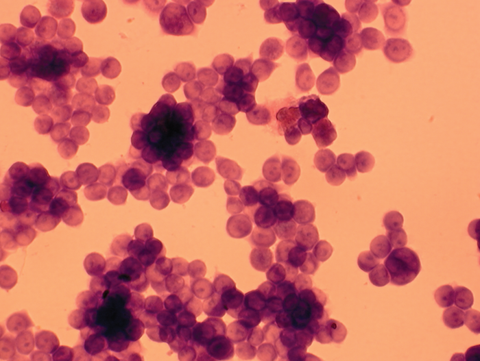
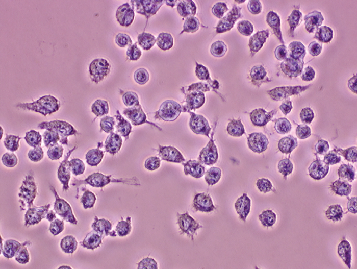
MORPHOLOGY OF CANCER CELLS
When normal cells are grown in culture they stop growing when they become overcrowded. This is called contact inhibition. Cancer cells in culture grow in an uncontrolled way because they have lost this property. This help tumors to form in the body. In addition, many different cell types can be present in a single tumor.
This experiment allows students to see the differences between normal and cancer cells in both their growth and cell types.
> Kit includes:
Multispot slides (2 cell types each), fixing agent, eosin and FlashBlue™ stain, mounting medium, cover slips, transfer pipets, and immersion troughs.
> You need:
Microscope with minimum 400x magnification.
ANALYSIS OF MAMMALIAN CELL TYPES
Your students will be amazed at the differences they observe between various mammalian cell types and how these cell function. cell are fixed on microscope slides and students stain the cells on the slide to view morphological characteristics of cell types.
These cells are very safe for classroom use.
> Kit includes:
Multispot slides (4 cell types each), eosin and FlashBlue™ stain, mounting medium, cover slips, transfer pipettes, and immersion troughs.
> You need:
Microscope with 400x magnification.
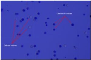
STUDY OF TOXICITY IN INSECT CELLS
Toxicity tests are of great importance to evaluate the safety of different compounds and prevent possible alterations that may be generated in living organisms, guaranteeing their health and well-being. One of the most used experimental approaches has been the use of cell cultures to evaluate the toxic potential of certain chemical products, drugs or pesticides. The objective of these practices is to introduce students to the experimental design of a toxicity study in cell cultures.
To ensure the success of the practice IT IS NECESSARY to coordinate the day of delivery of the kit with the day of the start of the practice. To schedule the delivery date of the kit, contact the person in charge of the cell culture laboratory at the email virgili@danagen.es
> Kit includes:
Insect cells (Sf9), culture and reaction medium, PBS, trypan blue, sterile flasks, culture plates, pipettes, scrapers, counting chambers, conical and centrifuge tubes.
> You need:
Microscope and inverted microscope, 70% ethanol, spray bottle, personal protective equipment, waste container, markers, micropipette and tips, automatic pipette or suction bulb.
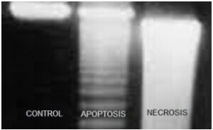
DETERMINATION OF APOPTOSIS BY DNA DETECTION
The term apoptosis describes a mechanism of programmed cell death in which cells self-destruct without activating inflammatory processes in response to cell death. Therefore, apoptosis is considered a physiological natural death process, which intervenes in the stages of elimination of unwanted cells. Death by apoptosis is evidenced in physiological processes such as the remodeling of bones and cartilage, during the morphogenesis of the fingers in the elimination of interdigital areas, during the elimination of the excess of neurons that is observed during the development of the nervous system, during organogenesis, etc. The objective of these practices is to offer students an alternative experimental tool in the identification of the process of cell death by apoptosis.
To ensure the success of the practice IT IS NECESSARY to coordinate the day of delivery of the kit with the day of the start of the practice. To schedule the delivery date of the kit, contact the person in charge of the cell culture laboratory at the email virgili@danagen.es
> Kit includes:
Insect cells (Sf9), culture and reaction medium, lysis buffer, culture flask, culture plates, pipettes, scrapers, conical and centrifuge tubes, counting chambers.
> You need:
Microscope and inverted microscope, agarose, bath, electrophoresis cuvette, centrifuge, 70% ethanol, isopropanol, aerosol bottle, personal protective equipment, waste container, markers, micropipette and tips, automatic pipette or suction bulb.
¡ NEW PROTOCOL !
 CEL-6
CEL-6
 6 groups of students
Download protocol: DETERMINATION APOPTOSIS DNA DETECTION
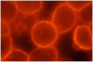
DETECTION OF CYTOSKELETON FIBERS THROUGH FLUORESCENCE
The cytoskeleton is a dynamic structure that allows the cell its movement, cell signaling, cell division, endocytosis, phagocytosis, communication between cell organelles, etc. The cytoskeleton is composed of different types of filamentous proteins, among which actin is found. There are two types of actin protein: depolymerized and polymerized or filamentous, involved in various cellular processes, being responsible for conferring the shape of the cell. The objective of these practices is to offer students an experimental tool for the detection of cytoskeleton fibers through the use of fluorescence.
To ensure the success of the practice IT IS NECESSARY to coordinate the day of delivery of the kit with the day of the start of the practice. To schedule the delivery date of the kit, contact the person in charge of the cell culture laboratory at the email virgili@danagen.es
> Kit includes:
Insect cells (Sf9), culture medium, fixation and permeabilization medium, coverslips and slides, mounting medium, PBS, culture flask, culture plates, pipettes, scrapers, conical tubes, counting chambers.
> You need:
Microscope and inverted microscope, fluorescence microscope (with filter for TRITC), 70% ethanol, isopropanol, aerosol bottle, personal protective equipment, waste container, markers, micropipette and tips, automatic pipette or suction bulb, tweezers and needles sleeveless.
top